
含细菌的肠绒毛
微生物组基础知识
微生物组基础知识

微生物组的组成
微生物组是指特定区域内微生物(微生物群)、其基因及其微环境(生活环境)的完整集合。细菌约占微生物组的 98%。1、2
微生物组的子集包括病毒组(病毒)、真菌组(真菌)和古生菌组(古生菌;这些微生物类似于细菌,但属于完全不同的域)。3微生物组是一个存在复杂相互作用和相互关联代谢作用的动态环境。4
病毒组包括会感染微生物组内细菌(噬菌体)以及环境中宿主细胞的病毒。5、6噬菌体占病毒组的大多数。5、6到目前为止,对病毒组的研究较为有限6、7,但是在患有慢性肠病或急性腹泻的犬体内,已确定了粪便病毒组的变化。5、6同样,目前已发表的关于宠物真菌组的研究也较为有限。8
肠道微生物组的基因数量远远超过宿主的基因数量。据估计,人体内微生物基因数量是人体基因数量的 100 倍以上。9
对幼猫肠道微生物组进行的宏基因组分析显示,发现的独特基因数量大约是猫基因组中已发现的开放阅读框数量的 108 倍。10
肠道微生物组中占比最多的菌门是厚壁菌门、拟杆菌门、梭杆菌门,其次是变形菌门和放线菌门。11
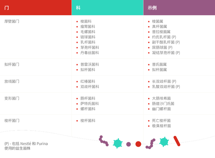
变形菌门是肠道微生物组中最多样化的菌门,包括许多已知的机会性致病菌(如 大肠杆菌、克雷伯氏菌、沙门氏菌和弯曲杆菌)以及在肠道内稳态中发挥重要作用的细菌。12猫粪便微生物组可能比犬粪便微生物组更为多样化。12
探索Microbiome Forum的其他部分


了解更多信息
- Barko, P.C., McMichael, M.A., Swanson, K.S., Williams, D.A. (2018). The gastrointestinal microbiome: a review. Journal of Veterinary Internal Medicine, 32, 9–25. doi: 10.1111/jvim.14875
- Marchesi, J. R. & Ravel, J. (2015). The vocabulary of microbiome research: a proposal. Microbiome, 3, 31. doi: 10.1186/s40168-015-0095-5
- Kim, J. Y., Whon, T. W., Lim, M. Y., Kim, Y. B., Kim, N., Kwo, M.-S.,…Na, Y.-D. (2020). The human gut archaeome: identification of diverse haloarchaea in Korean subjects. Microbiome, 8, 114. doi: 10.1186/s40168-020-00894-x
- Seth, E. C., & Taga, M. E. (2014). Nutrient cross-feeding in the microbial world. Frontiers in Microbiology, 5, 350. doi: 10.3389/fmicb.2014.00350
- Moreno, P. S., Wagner, J., Mansfield, C. S., Stevens, M., Gilkerson, J. R., & Kirkwood, C. D. (2017). Characterization of the canine faecal virome in healthy dogs and dogs with acute diarrhoea using shotgun metagenomics. PLoS ONE, 12(6), e0178433. doi: 10.1371/journal.pone.0178433
- Moreno, P. S., Wagner, J., Kirkwood, C. D., Gilkerson, J. R., & Mansfield, C. S. (2018). Characterization of the fecal virome in dogs with chronic enteropathy. Veterinary Microbiology, 221, 38–43. doi: 10.1016/j.vetmic.2018.05.020
- Zhang, W., Li, L., Deng, X., Kapusinszky, B., Pesavento, P. A., Delwart, E. (2014). Faecal virome of cats in an animal shelter. Journal of General Virology, 95, 2553–2564. doi: 10.1099/vir.0.069674-0
- Foster, M. L., Dowd, S. E., Stephenson, C., Steiner, J. M., & Suchodolski, J. S. (2013). Characterization of the fungal microbiome (mycobiome) in fecal samples from dogs. Veterinary Medicine International, 2013, 658373. doi: 10.1155/2013/658373
- Richards, P., Thornberry, N. A., & Pinto, S. (2021). The gut-brain axis: Identification of new therapeutic approaches for Type 2 diabetes, obesity, and related disorders. Molecular Metabolism, E pub ahead of print. doi: 10.1016/j.molmet.2021.101175
- Deusch, O., O’Flynn, C., Colyer, A., Morris, P., Allaway, D., Jones, P. G., & Swanson, K. S. (2014). Deep Illumina-based shotgun sequencing reveals dietary effects on the structure and function of the fecal microbiome of growing kittens. PLoS ONE, 9(7), e101021. doi: 10.1371/ journal.pone.0101021
- Suchodolski, J. S. (2011). The intestinal microbiota of dogs and cats: A bigger world than we thought. Veterinary Clinics of North America Small Animal Practice, 41, 261–272. doi: 10.1016/j.cvsm.2010.12.006
- Belas, A., Marques, C., & Pomba, C. (2020).The gut microbiome and antimicrobial resistance in companion animals. In Duarte, A. & Lopes da Costa, L. (Eds.), Advances in Animal Health, Medicine and Production (1st ed.), pp. 233–245. Springer International Publishing