
ANATOMICAL HEART
Cardiac Conditions
Cardiac Conditions

Cardiac disease is one of the most common disorders in dogs and cats.
Studies report that approximately 10% of dogs and 15% of cats have cardiac disease. The majority of cats suffer from cardiomyopathies. In dogs, the smaller breeds are most likely to have myxomatous mitral valve disease (MMVD) while larger breeds tend to develop dilated cardiomyopathy.1–4
Despite having heart disease, cats and dogs often appear healthy until their heart is failing. The early stages of disease may go undetected unless pets get diagnosed during a veterinary exam scheduled for a non-cardiac reason.1, 5, 6
The American College of Internal Veterinary Medicine (ACVIM) identifies four stages of heart disease in cats with cardiomyopathy or dogs with myxomatous mitral valve disease. Each stage is based on clinical and echocardiographic exams, and then linked with treatment targeted at managing clinical signs. Accurately identifying the stage of heart disease is important for treatment and prognosis.1, 7
Heart failure refers to the clinical signs that develop when the heart can no longer pump enough blood to meet the body’s needs. Once congestive heart failure (CHF) develops, pets have a significantly shortened life span.4, 8, 9
Read on to learn about research that may help veterinarians better predict heart disease progression or find new roles for nutrients in pets with heart disease.


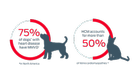
Most cats with cardiac disease have hypertrophic cardiomyopathy (HCM), a myocardial disease characterized by left ventricular hypertrophy that is not caused by other medical conditions. Without an echocardiogram, cats often escape detection in early stages of disease because they may not have a suspicious heart murmur or show clinical signs until they develop heart failure (CHF) or present with paralysis from aortic thromboembolism (ATE).4
Cats with HCM are more likely to be older, male, and have a loud systolic murmur. Most are also mixed-breed cats although some breeds, such as the Maine Coon and Ragdoll are at increased risk due to underlying genetic mutations.12
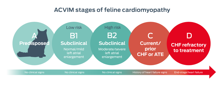
The presentation and outcome for feline cardiomyopathy is extremely variable. However, about 30% progress to heart failure. In general, the more severe the left atrial enlargement, the higher the risk for arterial thromboembolic episodes (ATE) and CHF.
Care for cats with CHF is based on managing clinical signs such as pulmonary edema and supporting heart function. Dietary recommendations focus on maintaining cats’ caloric intake, avoiding high sodium diets or treats, omega-3 fatty acid supplementation for heart health, and monitoring serum potassium levels.7,13


Like cats with early stage cardiomyopathy, dogs in early stage myxomatous mitral valve disease also appear healthy. MMVD is usually detected during a routine exam when a left apical systolic murmur is auscultated.1, 5
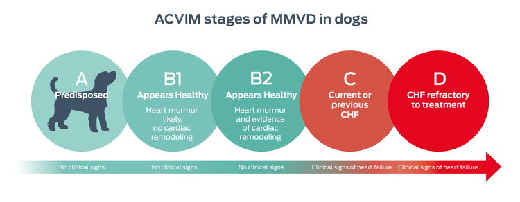
Progression of MMVD is also hard to predict, and left atrial enlargement is one of the most reliable indicators of advancing heart disease. Compared to dogs in early stage MMVD, those in congestive heart failure have shorter survival times.8, 14–16
Purina's research

Biomarkers can provide important clues about the progression of disease. In cardiac conditions such as MMVD, advancing stages are a risk for heart failure. Purina scientists studied microRNA in dogs with MMVD to assess the potential for new biomarkers.
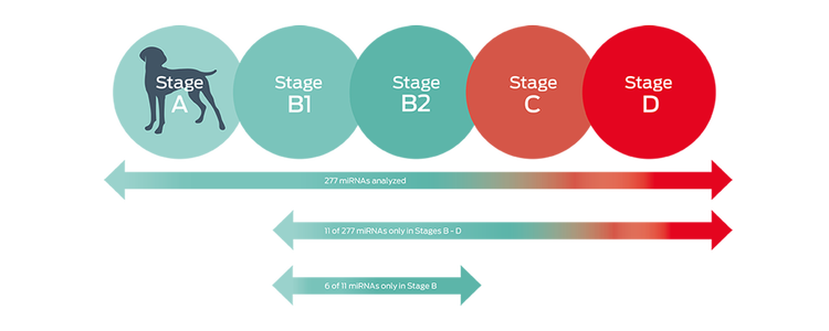
Purina scientists analyzed 277 circulating miRNA expression profiles from dogs in all stages of MMVD, from normal dogs to those in congestive heart failure.17
Although additional research is needed, the results suggest that some circulating miRNAs could be biomarkers for diagnosis, prognosis, or monitoring response to treatment in MMVD in dogs.
MicroRNAs (miRNAs) are small, non-coding RNA molecules that are potential noninvasive biomarkers for cardiac disease.
Using the ACVIM guidelines for staging dogs with MMVD, the researchers placed 18 dogs of various breeds into three groups of six dogs each.
Results showed that dogs in stages B, C, or D had 11 miRNAs that were differentially expressed compared to healthy dogs in stage A.
Among the 11 miRNAs that were differentially expressed, 6 miRNAs were significantly different between dogs in stage B1/B2 and those in stage C or D.
Gene expression changes were also greater as the severity of MMVD increased.
Key things to remember
- The American College of Veterinary Internal Medicine classification scheme has identified four stages of heart disease in cats with cardiomyopathies and in dogs with myxomatous mitral valve disease.
- Heart disease progression can be unpredictable and pets often appear normal in the early stages of heart disease.
- Finding new biomarkers for MMVD, such as miRNA, may improve early diagnosis for dogs with this heart disease.
Explore areas of transforming heart health:


Find out more
- Keene, B. W., Atkins, C. E., Bonagura, J. D., Fox, P. R., Häggström, J., Fuentes, V. L., Oyama, M. A., Rush, J. E., Stepien, R., & Uechi, M. (2019). ACVIM consensus guidelines for the diagnosis and treatment of myxomatous mitral valve disease in dogs. Journal of Veterinary Internal Medicine, 33(3), 1127–1140.
- Buchanan, J.W. Prevalence of cardiovascular disorders. In: Fox P.R, Sisson D.D, Moise N.S, editors. Textbook of Canine and Feline Cardiology: Principles and Clinical Practice. 2nd ed. Philadelphia, PA: WB Saunders; 1999. pp. 457–470.
- Payne, J. R., Brodbelt, D. C., & Luis Fuentes, V. (2015). Cardiomyopathy prevalence in 780 apparently healthy cats in rehoming centres (the CatScan study). Journal of Veterinary Cardiology: the official journal of the European Society of Veterinary Cardiology, 17 Suppl 1, S244–S257.
- Fox, P. R., Keene, B. W., Lamb, K., Schober, K. A., Chetboul, V., Luis Fuentes, V., … Tachika Ohara, V. Y. (2018). International collaborative study to assess cardiovascular risk and evaluate long-term health in cats with preclinical hypertrophic cardiomyopathy and apparently healthy cats: The REVEAL Study. Journal of Veterinary Internal Medicine, 32(3), 930–943.
- Côté, E., Edwards, N.J., Ettinger, S.J., Fuentes, V.L., MacDonald, K.A., Scansen, B.A., Sisson, D.D., & Abbott, J.A. (2015). Management of incidentally detected heart murmurs in dogs and cats. Journal of Veterinary Cardiology, 17(4), 245–261.
- Loughran, K. A., Rush, J. E., Rozanski, E. A., Oyama, M. A., Larouche-Lebel, É., & Kraus, M. S. (2019). The use of focused cardiac ultrasound to screen for occult heart disease in asymptomatic cats. Journal of Veterinary Internal Medicine, 33(5), 1892–1901.
- Luis Fuentes, V., Abbott, J., Chetboul, V., Côté, E., Fox, P. R., Häggström, J., Kittleson, M. D., Schober, K., & Stern, J. A. (2020). ACVIM consensus statement guidelines for the classification, diagnosis, and management of cardiomyopathies in cats. Journal of Veterinary Internal Medicine, 34(3), 1062–1077.
- Borgarelli, M., & Buchanan, J.W. (2012). Historical review, epidemiology and natural history of degenerative mitral valve disease. Journal of Veterinary Cardiology, 14(1), 93–101.
- Rush, J. E., Freeman, L. M., Fenollosa, N. K., & Brown, D. J. (2002). Population and survival characteristics of cats with hypertrophic cardiomyopathy: 260 cases (1990–1999). Journal of the American Veterinary Medical Association, 220(2), 202–207.
- MacDonald, K. Feline cardiomyopathy. In: Smith, F.W.K., Tilley, L.P., Oyama, M.A., & Sleeper, M.M, editors. Manual of Canine and Feline Cardiology. 5th ed. Saint Louis, MO: Elsevier; 2016. pp. 153.
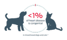
- Schrope, D. P. (2015). Prevalence of congenital heart disease in 76,301 mixed-breed dogs and 57,025 mixed-breed cats. Journal of Veterinary Cardiology: the official journal of the European Society of Veterinary Cardiology, 17(3), 192–202.
- Gil-Ortuño, C., Sebastián-Marcos, P., Sabater-Molina, M., Nicolas-Rocamora, E., Gimeno-Blanes, J. R., & Fernández Del Palacio, M. J. (2020). Genetics of feline hypertrophic cardiomyopathy. Clinical Genetics, 10.1111/cge.13743.
- Freeman, L. M. & Rush, J. Nutrition in Cardiovascular Disorders. In: Smith, F.W.K., Tilley, L.P., Oyama, M.A., & Sleeper, M.M, editors. Manual of Canine and Feline Cardiology. 5th ed. Saint Louis, MO.: Elsevier; 2016. Pp. 394–403.
- Ettinger, S. J., Benitz, A. M., Ericsson, G. F., Cifelli, S., Jernigan, A. D., Longhofer, S. L., Trimboli, W., & Hanson, P. D. (1998). Effects of enalapril maleate on survival of dogs with naturally acquired heart failure. The Long-Term Investigation of Veterinary Enalapril (LIVE) Study Group. Journal of the American Veterinary Medical Association, 213(11), 1573–1577.
- Häggström, J., Boswood, A., O'Grady, M., Jöns, O., Smith, S., Swift, S., … DiFruscia, R. (2008). Effect of pimobendan or benazepril hydrochloride on survival times in dogs with congestive heart failure caused by naturally occurring myxomatous mitral valve disease: the QUEST study. Journal of Veterinary Internal Medicine, 22(5), 1124–1135.
- Mattin, M. J., Boswood, A., Church, D. B., McGreevy, P. D., O'Neill, D. G., Thomson, P. C., & Brodbelt, D. C. (2015). Degenerative mitral valve disease: Survival of dogs attending primary-care practice in England. Preventive Veterinary Medicine, 122(4), 436–442.
- Li, Q., Freeman, L. M., Rush, J. E., & Laflamme, D. P. (2015). Expression Profiling of Circulating MicroRNAs in Canine Myxomatous Mitral Valve Disease. International Journal of Molecular Sciences, 16(6), 14098–14108.